Positron Emission Tomography (PET) Scans Lab
For this lab you should read the text below and the linked articles on PET-based research. Then go to Beachboard and take the quiz for the lab.
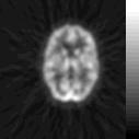
PET (Positron Emission Tomography) scans are non-invasive imaging technique used in cognitive science to study brain function and dysfunction. PET scans differ from some other imaging techniques in that PET scans allow cognitive scientists to observe brain functioning by providing measures of brain activity based upon metabolic activity.
Radioisotopes and PET
PET scans require the injection of a small amount of biologically relevant material like oxygen or glucose (sugar) which have been labeled with radio nuclides such as carbon-11, nitrogen-13, oxygen-15 and fluorine-18 (fluorine-18 is the most common). To understand the function of isotopes in PET scans, let's talk briefly about radiation in general.
While working with uranium containing compounds in 1896, Henri Becquerel made an unexpected discovery: Covered photographic plates became partially exposed in the presence of his compounds. Becquerel hypothesized that the exposure was due to some kind of ray passing through the plate coverings. He went on to discover several materials other materials in addition to uranium that also emitted these rays. Materials that emit this kind of radiation are said to be radioactive and to undergo radioactive decay. Ernest Rutherford performed experiments in 1899 to determine that uranium compounds produce three distinct kinds of radiation. Rutherford used the penetrating abilities of each of these radiations to differentiate and name them. The first, α (alpha) radiation proved the weakest form of radiation. A sheet of paper can stop α- radiation, which Rutherford determined was the nucleus of a Helium (He) atom. The next strongest form of radiation is β (beta) radiation. β-radiation turns out to be electrons at high velocity, and one needs 6mm of aluminum to stop β particles. Finally, the most powerful radiation is γ (gamma) radiation. γ rays are high energy photons that one needs several millimeters of lead to stop. Gamma rays are thus a form of electromagnetic radiation, just like visible light or X-rays. However, γ rays have a much shorter wavelength and a much higher energy than visible light. Even X-rays with the shortest wavelength and highest energy only overlap into the the range of "long"-wavelength (lower energy) gamma rays. The distinction between X-rays and γ rays is not merely their energy, but also the source of the respective radiations. X-ray photons are generated by energetic electron processes and not nuclear decay. The gamma ray spectrum is usually defined as light having a frequency between 1018 and 1021 Hertz. All radiation, as you might have guessed, is the result of atoms losing some particle or particles. This process of atomic degeneration is radioactive decay.
 |
α-particle radiation is a Helium nucleus (two protons and two neutrons) produced by nuclear fission. A massive nucleus separates into two less-massive nuclei (one of them the alpha particle). |
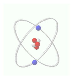 |
β-particle radiation is an electron that emerges from the decay of one of the neutrons inside an atom into a proton, the β-electron and an anti-electron-type (positron) neutrino. |
 |
A gamma particle is a photon. It is produced as a step in a radioactive decay chain when a massive nucleus produced by fission relaxes from the excited state in which it first formed towards its lowest energy or ground-state configuration. In PET scanning γ-radiation is released when a positron leaves the nucleus during radioactive decay (see above) and collides with an electron. |
Returning to isotopes, an important property of atoms is their atomic number, which physicists define as the number of protons in the nucleus and denote with the symbol Z. An atom's atomic number determines its chemical properties. Similarly, an atom's atomic mass number is symbolized by A, and is defined as the total number of nucleons (protons and neutrons) in the atom. Isotopes are atoms with identical atomic numbers but different atomic masses. Though isotopes have the same chemical properties as other atoms of their element, they have very different nuclear properties. For example, though many isotopes are stable, some isotopes have unstable nuclei and are radioactive. Hydrogen atoms normally have one neutron and no protons. Tritium (one proton and two neutrons) is a radioactive isotope of hydrogen, while deuterium (one proton and two neutrons), another isotope of hydrogen is stable. The isotopes used in PET scans are all radioactive and decay rapidly. Commonly used isotopes include: Carbon-11 or 11C is a radioactive isotope of carbon that decays 100% into Boron-11 by positron emission. Its half-life, or the time it takes half of the Carbon-11 to decay into Boron-11 is approximately 20 min. Nitrogen-13 or 13N is a isotope of nitrogen having a half life of approximately ten minutes which is tags ammonia. Oxygen-15 or 15O is an isotope of oxygen having half-life of approximately 2 min by the emission of positrons. Perhaps the most common isotope used in PET scans of the brain is Fluorine-18 or 18F, which is a fluorine isotope having a half-life of approximately 110 minutes. Fluorine-18 is very useful because of its long half-life, and because it decays be emitting positrons having the lowest positron energy, which generates allows for the sharpest images with a high-resolution PET.
The radioisotopes attached to the glucose or oxygen cause the glucose or oxygen to emit positrons. Once introduced into the body, organs and tissues process these radioactive agents as part of their normal metabolic function. For instance, since brain cells need sugar in the form of glucose to operate; the more they operate, the more glucose they require. Thus, the radioactive glucose injected by the cognitive scientist travels to the brain, where it collects in greater concentration in the most active areas of the brain. The PET scanner detects the location and concentration of the radioactive glucose or oxygen within the various areas of the brain by detecting the gamma rays given off when the radioactive labeled glucose or oxygen emits positrons (i.e., during radioactive decay).
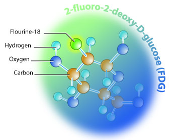 |
| A diagram of 2-fluoro-2-deoxy-D-glucose or
FDG, a radiolabeled glucose atom used in PET scans
Diagram courtesy of Bioteach at UBC |
The PET Scan Process
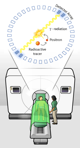
In a typical case, subjects are injected with glucose or inhale oxygen, and are then placed on a flat table. The table moves in increments through a "donut" shaped housing (see below) which contains the sensor array. As the isotope used in the radiolabeled oxygen or glucose undergoes radioactive decay, it emits high energy positions (anti-matter) which collide with electrons resulting in bursts of γ-radiation. The detector array within the PET scanner detects the γ-rays resulting from the positron/electron collisions as they are emitted from a thin slice of brain with each increment.
 |
 |
|
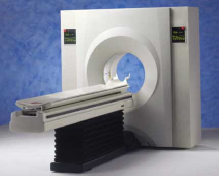
Image of PET Scanner Image from Riologyinfo |
Diagram of PET Scan Process Diagram courtesy of Bioteach at UBC |
The
detected signals are then processed to extrapolate the exact origin of each γ-ray.
The result is rendered, often using colors (see below) to highlight the different levels of
activity (i.e., where more glucose or oxygen is going), a computer creates a picture of the activity.
The table is then moved and the process repeated, resulting in a series of thin slice images of the
bodily region of interest (e.g. brain, breast, liver). This creates a series of thin slice images,
which can be assembled into a three dimensional representation of the total area
of interest. The resolution of PET Scan images allow scientists to study brain activity in structures as small as 1/5" x 1/5".
One drawback for PET scans is that PET centers must be located near a particle accelerator device that produces the short-lived radioisotopes used in the technique. Another drawback, of course, is that the technique involves exposing people to radiation, though the level is only about as high as exposure due to taking a cross-country air flight.
Single Photon Emission Computerized Tomography (SPECT) is a similar tomographic technique. However, the substances used in SPECT are radioactive labeled with isotopes having longer decay times than those used in PET. This makes SPECT centers more common and SPECT scan much cheaper. However, the radio labeled substances used in SPECT emit single instead of double gamma rays. Thus, though SPECT can provide information about blood flow and the distribution of radioactive substances, SPECT images have less sensitivity and can only image larger structures (about 1/2" by 1/2") with less detail. Note that there are about 10,000,000 neurons per square centimeter of cortical surface.
The Subtraction Technique for using PET Scans for Complex Tasks
In many cases the tasks that cognitive scientists which to study--the target or primary task--also require that the brain perform other tasks--the secondary tasks. For example, the reasoning tasks we looked at during the logic lectures involved not only deductive or inductive reasoning tasks (primary or target), but also language comprehension tasks as well (secondary). In order to better isolate the areas of the brain involved in one task when the brain may have to perform several tasks, scientists perform PET scans on subjects during the performance of two tasks different tasks. The first task will represent the other tasks hypothsized to be operant (the secondary tasks) with the exception of the task of primary interest (target). For instance, in the reasoning studies done by Osherson et al. (1998) subjects performed both reasoning tasks and meaning tasks. Since language comprehension was clearly involved in evaluating the arguments, subjects were asked to peform the meaning tasks. As Osherson et al. express it, "The meaning task required subjects to examine premises and conclusion individually and determine whether any had anomalous content; it served as a baseline condition since no more than sentence comprehension was involved." (1998, p. 370) The second task is the target task--the task of primary interest incuding the necessary secondary tasks. In the case of Osherson et al. subjects evaluated whether arguments were valid or invalid in the deductive logic task. Scientists then subtract the activation patterns in the performance of the secondary only task from the activation patterns during the target task (combined primary and secondary task). The remaining activation is then hypothesized to result exclusively from the primary or target task and not the secondary tasks.
Read These Articles About Pet Scan Discoveries Before Doing The Quiz!!!
Methamphetamine Withdrawal Is Associated With Brain Changes
Some Great General PET Links: